
FAQs About X-Rays


X-ray imaging is a commonly used tool in the diagnosis of X-linked hypophosphatemia (XLH), a rare genetic disorder characterized by low phosphate levels in the blood.
This article answers some of the frequently asked questions (FAQs) about X-rays and how they can be useful in diagnosing XLH.
What are X-rays?
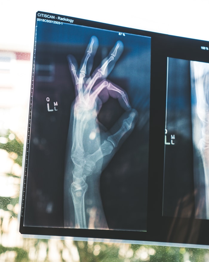
X-rays are electromagnetic waves, a type of radiation that can penetrate the body and are reflected back by hard tissues like bones and cartilage. The reflected X-rays are captured on a film, which helps in studying the structure of bones.
How does X-ray imaging help in XLH diagnosis?
XLH is characterized by rickets-like symptoms such as osteomalacia, or soft and weak bones, early osteoarthritis, fractures, pain, dental abscesses, or a buildup of pus in the teeth, and stunted growth. All these symptoms can be observed in detail via X-ray imaging. The results of X-ray imaging can be corroborated by other tests to confirm an XLH diagnosis.
Are X-rays safe?
The amount of radiation received during a typical X-ray examination is smaller than what is generally received from natural sources every day. The radiation dose is matched to adhere to defined safety criteria. X-rays also are non-radioactive. Therefore, adverse effects are extremely rare.
How should I prepare for an X-ray session?
The required preparation for a typical X-ray session is minimal and, in most cases, just involves changing into a gown. Chains, watches, rings, and any other objects that can potentially show-up extraneously on the X-ray film need to be removed before the test.
There usually are no food restrictions unless the procedure involves the administration of a contrasting agent. But this is usually not necessary when XLH is suspected.
Who will interpret my X-ray report?
Although an X-ray exam is done by a qualified medical radiation technologist (MRT), that individual is only eligible to determine the quality of the output image. The MRT can not interpret the results.
An X-ray report, based on the output image, will be prepared by a radiologist, who is a doctor specially trained to interpret radiological scans. Usually, this report provides enough information for the patient’s own doctor to form an opinion.
How long will the results take to arrive?
Most X-ray reports are made available within two to three business days. However, a faster turnaround may be possible in cases of urgency, or upon request by your doctor.
Can I undergo X-ray imaging during pregnancy?
Yes. Inform your doctor and the MRT beforehand so that certain precautions can be taken, such as shielding the pelvic region, to minimize the exposure of the fetus to radiation. If imaging the pelvic region is necessary, the MRT usually ensures that the radiation dose is kept as low as possible.
Can I decline an X-ray investigation?
X-ray imaging provides vital clues in understanding the progression of the disease. If, for some reason you decide not to undergo an X-ray exam, be sure to inform your doctor so that an alternative diagnostic procedure can be prescribed.
Last updated: Feb. 07, 2020.
***
XLH News Today is strictly a news and information website about the disease. It does not provide medical advice, diagnosis, or treatment. This content is not intended to be a substitute for professional medical advice, diagnosis, or treatment. Always seek the advice of your physician or other qualified health provider with any questions you may have regarding a medical condition. Never disregard professional medical advice or delay in seeking it because of something you have read on this website.